It doesn’t take a brain surgeon to know that medicine is changing every day. But for most of us, it’s not as obvious just how quickly some of these advancements in neurology and neurosurgery are improving medical care. These breakthroughs make it seem like we’re living in the future. Because, well, we kind of are.
AI Technology

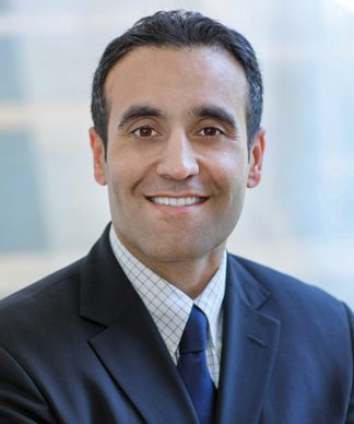
Navid Redjal, MD, director of Neurosurgical Oncology at Capital Health
The brain is not just a part of your body – it coordinates the movements, functions and feelings that determine who you are. So that makes personalizing treatments much more important, says Navid Redjal, MD, director of Neurosurgical Oncology at Capital Health.
The latest AI technology allows neurosurgeons to get the most accurate map of the brain and spine before heading into surgery. The data helps them see hidden patterns in brain activity, giving a better understanding of how the brain operates to create a personalized surgical plan.
“We can read data that was very difficult to decipher before to personalize surgery in a way we never could have without AI,” says Redjal.
Tumors that may once have been deemed inoperable are suddenly accessible because doctors can see exactly how to remove the tumor without damaging brain functioning. “It leads to better quality of life and safer surgeries,” Redjal says.
Treatable Dementia

Patrick Connolly, MD, chief of neurosurgery at Virtua Memorial Hospital
When an older adult experiences cognitive decline, dementia is often the concern. But that’s not always the cause, says Patrick Connolly, MD, chief of neurosurgery at Virtua Memorial Hospital.
As many as 10% of people diagnosed with dementia may actually have abnormal buildup of fluid in the brain, which is a more treatable condition called normal pressure hydrocephalus, says Connolly.
There are 3 main symptoms that, when experienced together, are telltale signs someone may have NPH – trouble walking that mimics Parkinson’s disease, cognitive impairment and urinary incontinence.
Diagnosis is fairly straightforward, says Connolly. Doctors can perform a spinal tap, where they’ll draw a few tablespoons of spinal fluid – a noninvasive outpatient procedure – to see if the patient’s symptoms get better. If the symptoms ease, they may be able to get a more definitive treatment involving a shunt, which diverts the fluid out of the brain through a tube that runs under the skin into the stomach.
“It’s not guaranteed to work – around 80% of patients who see relief from the spinal tap will respond well to the shunt,” says Connolly. “But for those who do, the shunt can last 5 to 10 years before symptoms return in earnest.”
3D-Guided Imagery

Hekmat Zarzour, MD, neurosurgeon at Jefferson Health – New Jersey
In neurosurgery, going a millimeter in the wrong direction during surgery can have catastrophic outcomes. Now with 3D-guided imagery, doctors can see the brain and spine from every angle and avoid going too far. “With 3D imaging, we can rotate the images to see every aspect of the tumor. We can also see the vessels around it in a way that we never could have before,” says Hekmat Zarzour, MD, a neurosurgeon at Jefferson Health – New Jersey.
To get the image, doctors secure the patient’s head so it cannot move, attach antennas and use a machine to scan the face – resulting in a 3D roadmap that helps them know what to expect before surgery. Doctors can now be more aggressive because they can see small tumors that, without such detail, would not be accessible.
Take, for example, a tumor in the cerebellum, which controls fine motor skills.
“Before, it may have been too risky to operate,” says Zarzour. “Now we can clearly see the edges of the tumor, accurately measure how to get there and treat it with minimal damage.”
Stroke Treatment Breakthrough

Tudor Jovin, MD, medical director of Cooper Health Neurological Institute
A new study may change the way doctors all over the world treat strokes.
“After 5 years of clinical trials, we were able to prove, definitively, that a minimally invasive technique known as a mechanical thrombectomy is the best way to treat strokes in the back of the brain,” says Tudor Jovin, MD, medical director of Cooper Health Neurological Institute.
Jovin was among 5 doctors to recently present the study to the European Stroke Organization. This technique has long been used for strokes caused by blood restrictions in the front of the brain. But before, there wasn’t enough evidence to say it worked for strokes in the back of the brain, which are less common but significantly more dangerous, says Jovin.
“It’s likely this is going to become the new guideline for hospitals across the world,” he adds. “And it’s going to save a lot of lives.”
Robotic Navigation

R.J. Meagher, MD, neurosurgeon at Inspira Health
No, robots don’t perform brain and spine surgery. But they can assist, says R.J. Meagher, MD, a neurosurgeon at Inspira Health. “A major development in neurosurgery is a robot-assisted navigation,” says Meagher. “Using this platform, the robot identifies and navigates where it needs to go.”
Surgeons pick a target based on a preoperative image, and the robot can act as an arm to go where surgeons need it to go during spinal surgery – creating an even more minimally invasive surgery with higher accuracy, he says.
Because the robot is navigating, it also means patients don’t have to be repositioned during surgery, which can be risky. This cuts surgical time dramatically, says Meagher.
“Using this tool,” he says, “surgeons can do what we’ve been doing for years – more accurately and safely than ever before.”
Brain Cell Connections

Sarah West, PhD, neuropsychologist with Bancroft NeuroRehab
In the brain, there’s much more going on than meets the eye (or the MRI), says Sarah West, PhD, a neuropsychologist with Bancroft NeuroRehab. And now there are ways to observe the brain that help fine tune treatments for head injuries.
Deep Probabilistic Imaging (DPI) captures brain cell connections, as opposed to a routine MRI or CAT scan that captures a photo of the brain itself. This allows doctors to observe and treat injuries that you can’t physically see on the brain.
“If there’s a tumor or a lesion, you can see that,” says West. “But when brain cells aren’t connecting the way they should, it’s not always immediately visible. Think of it this way – if you’re in a helicopter, you can take a photo of a forest and zoom in as much as you want, but it’s not going to show you the broken roots under the soil.”
This imaging is especially impactful for injuries like a concussion or stroke, which can damage neuropathways. Now doctors can see exactly where these symptoms are coming from and develop a treatment plan.
“Even the idea of acknowledging that something is wrong can be really helpful for patients,” says West. “Patients can say they’re having trouble planning, organizing, problem-solving, thinking quickly, memory – whatever it is – but their CAT scans and MRI come back completely fine. That’s extremely disheartening because they know there is something wrong.”
This DPI imagining becomes tangible proof of what the patient is experiencing, which in itself can be a relief to them, she says.
“It allows doctors to jump into action much more quickly because they can now see the problem, and they can send proof to insurance companies that may not have covered treatment without it,” says West. “We can avoid a delay in treatment that could ultimately make the issue worse.”
“Broken brain cell connections can impact every part of who you are and how you function,” says West. “This allows us to increase a patient’s quality of life much more quickly and efficiently than we could before.”


